Living multispectral fluorescence imaging
Foreword
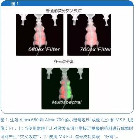
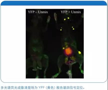
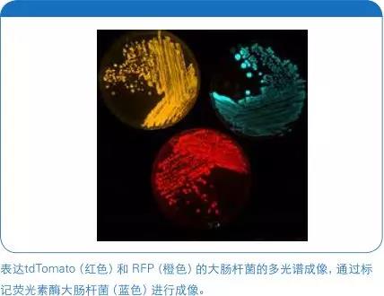
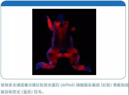
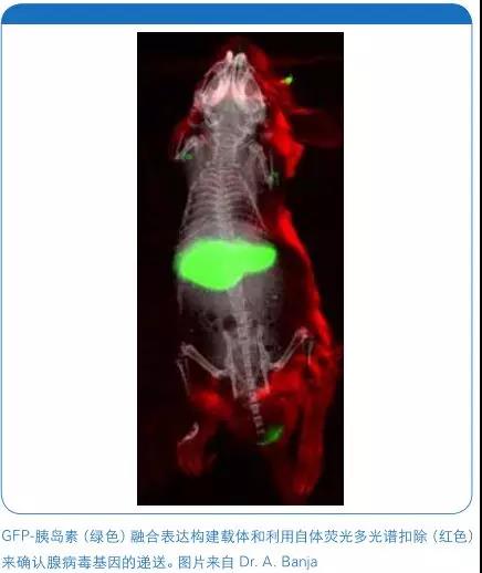
Conventional living body optical fluorescence imaging (FLI) using an excitation filter and an emission filter. This has many limitations for distinguishing between targeted signals, possible reporter signals, and autofluorescence tissue signals. Multi-spectral (MS) FLI using multiple excitation filter and a single emission filter, the excitation filter with a single or a plurality of emission filters, can produce a unique fluorescence spectrum curve of the material or region. (1) Therefore, the image of each fluorescent component pixel may thus be confirmed (FIG. 1).
Why choose live multispectral fluorescence imaging?
There are two main reasons for choosing a live MS FLI:
When 1- shorter wavelength for imaging reporter gene within a particular range of tissue autofluorescence can be suppressed autofluorescence background signal.
2- Imaging with a longer wavelength reporter gene (eg NIR) with overlapping spectra
At the same time, cross-reactivity can be suppressed to achieve clear detection of unique reporter genes.
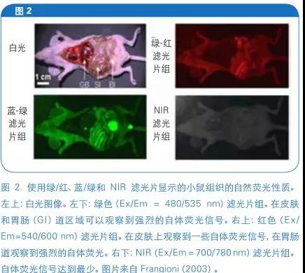
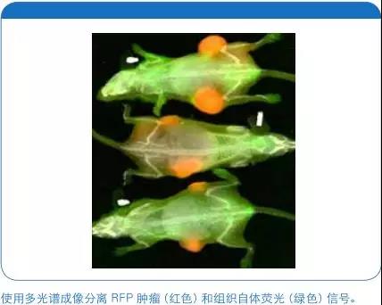
In order to understand these two reasons, it is first necessary to understand the basic knowledge of mouse tissue autofluorescence and its imaging in different ranges of fluorescence spectra. Figure 2 below shows the typical autofluorescence characteristics of mouse tissue when using the green/red, blue/green filter and NIR filter combination (2). It is worth noting that the green/red and blue/green filters exhibit higher levels of tissue autofluorescence, but NIR filters exhibit relatively less tissue autofluorescence. The use of MSFLI in the use of reporter genes such as green fluorescent protein (GFP), red fluorescent protein (RFP), fluorescein isothiocyanate (FITC) and Alexa 600 can effectively reduce tissue autofluorescence. Far red (650-700 nm) or near-infrared (NIR; 700-900 nm) fluorophores are used as an alternative because skin autofluorescence is small in this spectral range, but multiple filters are required (3). This method can simultaneously detect multiple molecular markers, such as fluorescent cell tracer reporter probes and fluorescent protease activity reporter probes. In addition, rodent foods often contain chlorophyll-containing plant material that may produce strong autofluorescence in the low NIR spectrum. Multispectral imaging is often used to suppress this gastrointestinal (GI) autofluorescence, avoiding the need to replace the feed with a purpura-free murine feed for imaging studies.
Start up and collect
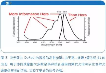
The Brooke system uses a "spectral-based" multispectral imaging approach. Wherein the plurality of excitation filters and a single emission filter is used to capture the "superimposed image." For most organic fluorophores include fluorescent protein gene including the excitation spectrum can provide more information than an emission spectrum (FIG. 3), based on the transmitted multi-spectral imaging compared, based on multispectral excitation imaging can better capture these different information.
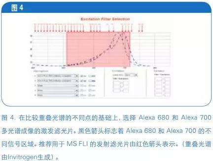
A total of 28 excitation filters are configured in the system. The optimal excitation filter should be selected for a given imaging experiment before defining the emission filter. In order to better select the filter, it is very useful to compare the fluorescence spectrum difference of the excitation spectrum curve of the reporter gene used for imaging in advance . In the example below (Fig. 4) Alexa 680 and Alexa 700 , there is a significant difference in the excitation spectra between the two at 520 and 720 nm. (A solution of Alexa 680 and Alexa 700 complete content filter selection and modeling, check the webinar: https://youtu.be/iEyLl3qAzpA).
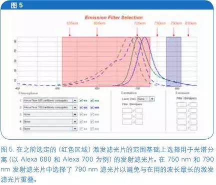
The system is configured with six emission filters (535 nm, 600 nm, 700 nm, 750 nm, 790 nm, and 830 nm). Emission filters that do not overlap within 60 nm of the longest wavelength excitation filter are typically selected . Emission filter need not be uniform with the selected fluorescent emission peak, the filter should be preferred to cover the excitation light filter sheet specific fluorescence spectra. Alexa 680 and 700 continue to be imaged, for example, 535,600,700 and 750 nm due to the bandwidth of the filter with the selected excitation filter range (FIG. 5) overlap, so there may be selected a filter 790 nm .
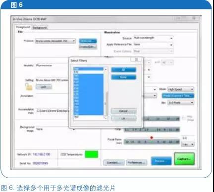
In "capture (Capture)" dialog box (FIG. 6) to select a plurality of support pieces for acquiring programming excitation filter. Multispectral acquisition should be performed as part of a post-multispectral modeling and/or separation experimental approach.
Optimization and multispectral modeling
The initial imaging and research setup includes initial steps for optimizing setup and modeling:
1-fluorophore imaging (in vitro)
2- Generate a spectral model
3- In vivo model evaluation
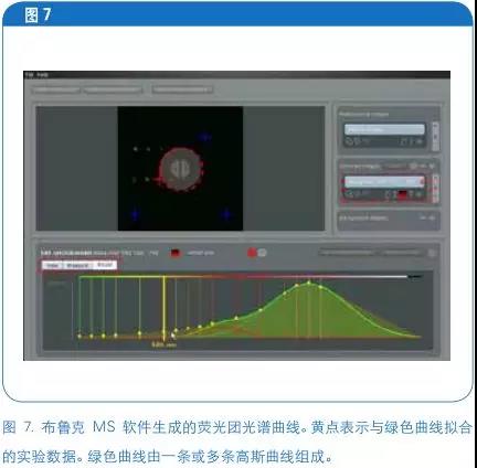
First, we recommend that you image the diluted fluorophore using the filters identified above . Once the image was acquired, a spectral curve was created by fitting a Gaussian curve to the experimental curve of the fluorophore (Figure 7).
Applied spectral model
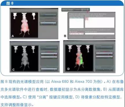
Once the data set to achieve the optimal spectral curve, the model can be called directly into the following years, without modification. For an existing model to the new data set, then the choice of the "isolated image" panel "+", and select the required model "isolated (UNMIX)" button (see FIG. 8). The system assigns the model to each pixel (4) using a least squares method suitable for solving multispectral models . To this end, the sum of the spectra in each pixel needs to match the sum of all possible combinations in the reference library. The separation algorithm also adds some additional restrictions (such as non-negative). Therefore, the key to successful spectral separation is to obtain an accurate spectrum of the library fluorophore. Once the spectral effects in each fluorophore are determined , the acquired superimposed image can be separated into separate images of the individual fluorophores .
Each "separated" image consists of overlapping images from separate "channels" ( using the Alexa 680 and Alexa 700 above). Individual images can be opened in the Bruce MI software and analyzed using relevant data from the Region of Interest (ROI) application , such as average, sum and net light intensity, area and perimeter, and automatic ROI lookup function and image math. In other words, the strength of the camera may be released and measured light-emitting substance animal "true" signal strength correlated in a complex manner. After the sensor to capture the signal, then down the multispectral analysis can produce an accurate quantitative data of the specific component (1).
Multispectral imaging in infectious disease, oncology and nanoparticle tracer studies
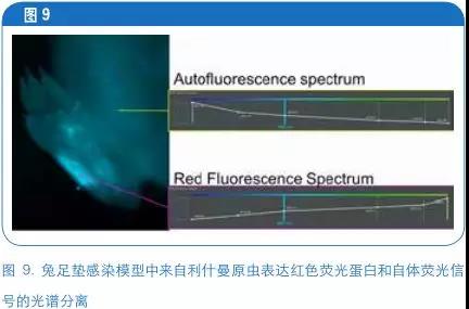
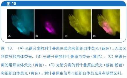
Fluorescence spectroscopy studies can be modeled to benefit from even more NIR fluorophores using green or red or fluorescent reporter gene. Multi-spectral imaging can be used to identify and model different spectral profiles of reporter genes, thereby reducing autofluorescence. 9 and 10 show the collected tissue autofluorescence deducted Leishmania infected footpad model rabbit imaging (M. Leevy, University of Notre Dame, unpublished) with the multi-spectral method.
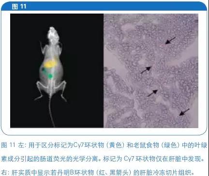
In another example, multispectral imaging was used for an in vivo polymer particle tracing study (5). Four hours after injection, in the region of the liver (FIG. 11) detects the particle Cy7 labeled crosstalk signals may be generated by the food in the GI chlorophyll obtained is isolated region, providing a clear signal to the positioning particles.
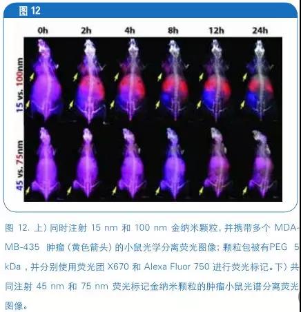
In a nanoparticle/tumor study, nanoparticle biodistributions of different sizes were detected in the same tumor mouse (6). Nanoparticles are differentially labeled, multi-spectral imaging is also used to signal (FIG. 12) separation cycle nanoparticles.
Release all the potential of in vivo fluorescence imaging

to sum up
In vivo multispectral fluorescence imaging can deduct tissue autofluorescence and perform multiple fluorophore imaging. This can enhance the signal to noise ratio and advanced multiple fluorescence imaging, to achieve more powerful study design.
references
[1] Levenson RM, Lynch DT, Kobayashi H, Backer JM, Backer MV (2008). Multiplexing with multispectral imaging: from mice to microscopy. ILAR J 49-78.
[2] Frangioni, JV (2003). In vivo near-infrared fluorescence imaging. Curr Opin Chem Biol.;7:626-34.
[3] Weissleder R, Ntziachristos V (2003). Shedding light onto live molecular targets. Nat Med. 9( 1 ) 123-8.
[4] Farkas DL, Du C, Fisher GW, Lau C, Niu W, Wachman ES, Levenson RM (1998). Non-invasive image acquisition and advanced processing in optical bioimaging. Comput Med Imaging Graph. 22(2):89 -102.
[5] Alexander L, Dhaliwal K, Simpson J, Bradley M. (2008) Dunking doughnuts into cells--selective cellular
Translocation and in vivo analysis of polymeric micro-doughnuts. Chem Commun (Camb). 14;( 30 ):3507-9
[6] Chou LY, Chan WC (2012). Fluorescence-tagged gold nanoparticles for rapidly characterizing the size-dependent biodistribution in tumor models. Unpublished.
Venlo structure is a popular one at present. This architecture uses horizontal girder as main bearer, forming a stable structure with columns. There is consolidation between horizontal girder and column, and hinge is used to connect column and foundation.
In Venlo greenhouse, foundation is made of reinforced concrete and the side wall is made of brick or reinforced concrete plate. The steel frame always use hot dip galvanized light steel. Roof beam adopts horizontal girder structure and using herringbone connection. The horizontal girder bears 2 or more roofs, which is made of aluminum alloy. This material is used as roof structure material and also glass inlay material. Other beams using gutter style to minimize the section. Delighting material of the roof and side wall using the hollow double-layer or multilayer PC board.
PC board is mainly made of PC/PET/PMMA/PP materials, it need sun screen coating to resist ultraviolet resist and ageing, and it is also required to be anti condensation and antidrug.
As a kind of light material, use PC board can greatly reduce the weight of greenhouse structure, and corresponding reduced the size of the steel frame, which saving the cost of steel.
Pc Board Venlo Greenhouse,Venlo Greenhouse,Venlo Type Pc Sheet Greenhouse
JIANGSU SKYPLAN GREENHOUSE TECHNOLOGY CO.,LTD , https://www.spgreenhouse.com