About phage culture and application
A1: Phage display technology is a method for cloning a gene encoding a polypeptide or a protein or a gene fragment of interest into a phage coat protein structural gene, and when the reading frame is correct and does not affect the normal function of other coat proteins, The exogenous polypeptide or protein is expressed in fusion with the coat protein, and the fusion protein is displayed on the surface of the phage with reassembly of the progeny phage. The displayed polypeptide or protein can maintain a relatively independent spatial structure and biological activity to facilitate recognition and binding of the target molecule. After the peptide library and the target protein molecule on the solid phase are incubated for a certain period of time, the unbound free phage is washed away, and then the phage which is bound to the target molecule is adsorbed by the competitive receptor or acid, and the eluted phage infects the host cell. After propagation and amplification, the next round of elution is carried out. After 3 to 5 rounds of "adsorption-elution-amplification", the phage specifically binding to the target molecule is highly enriched. The resulting phage preparation can be used to further enrich the target phage with the desired binding properties.
Q2: What are the advantages of phage display compared to other antibody development techniques?
A2: Compared with hybridoma technology, phage display technology has great advantages. The hybridoma method is only applicable to mice, rats, hamsters and guinea pigs. On the other hand, phage display can be used for all popular antibody-producing species including, but not limited to, humans, mice, rats, rabbits, chickens, camels, camels, alpacas, cattle, dogs, sheep, monkeys and sharks. High affinity monoclonal antibodies. Hybridoma-based monoclonal antibodies are developed to produce only a small amount of antibodies against a particular immunogen, while phage display technology can present an entire antibody repertoire of immunized animals, with almost 10% of antibodies being immunogen specific. The chances of finding antibodies with the desired properties using phage display technology are much greater. Furthermore, using hybridoma technology, it is difficult to combine an enrichment step that is capable of selectively isolating antibodies having the desired function. In most cases, all hybridoma clones are first generated and then verified one by one. In contrast, phage display technology allows for various enrichment strategies: antibodies with the desired properties can be enriched so that antibodies that do not have the desired function can be excluded from further validation. For example, antibody library screening can use a target to capture a strong conjugate while using control to block/deplete the cross-reacting conjugate. In summary, this method of immunosorbent library allows for the collection of almost all antibodies in an animal and the separation of the strongest conjugate from the collection.
Q3: What is the difference between the M13 , T7 and T4 phage series?
A3: M13 filamentous phage has been the most popular choice and is widely used in various types of research. The viral envelope consists of five distinct capsid proteins, including a major capsid pVIII (2,700 copies) and four minor capsids (pIII and pVI at one end and pVII and pIX at the other). Unlike T4 and T7, M13 is a lysogenic phage that assembles in the periplasm and is secreted from the bacterial membrane without lysing the host. Multiple capsid proteins on M13 phage provide a comprehensive display of choice for a wide range of peptides and proteins with unique properties. These five capsids have been successfully used for display in the outer domain with a unique carrier. PIII and PVIII are the primary choices for M13 phage display.
Phage T4 differs from M13 in many respects. T4 has a larger size, a tail structure and a double-stranded DNA (dsDNA) genome encoding 50 different proteins, and larger genomic DNA allows for the insertion of larger foreign proteins. Two different domains, two non-essential coat proteins, can be displayed on the HOC and SOC. N- and C-terminal insertions can be used. In the absence of a membrane secretion process, host toxicity can be avoided using T4 phage.
Compared to filamentous phage and lambda phage, T7 phage has a shorter life cycle. The assembly of the progeny phage is released in the bacterial cytoplasm by cell membrane lysis, so there is no host toxicity due to size limitation and secretion process. In addition, T7 phage is very stable under extreme conditions in which other phage cannot survive.
Q4: Can I construct a cDNA library using the M13 phage display system?
A4: Usually M13 is not suitable for cDNA expression because M13 phage display requires in-frame expression between the leader sequence (required for secretion) and the N-terminus of the coat protein pill or pVIII. In order to properly fuse the corresponding protein into the coat protein, the insert must be in the correct reading frame at both ends and does not contain an in-frame stop codon. This results in very few clones produced in the M13 cDNA library.
Q5: What medium can be used to culture phage?
A5: TSB / TSA can be used to culture most phage. However, for different phage display systems, suitable media can be varied.
Q6: Which E. coli can be infected with phage M13? Does it have the opportunity to contaminate all cell lines in our laboratory?
A6: M13 phage is a filamentous phage that uses the tip of the bacterial F-binding pili as a receptor to promote the infection process. Therefore, they are specific only to E. coli strains containing the F plasmid (F+). For strains in the laboratory, we recommend that you check if they contain the F plasmid. If they do not, M13 phage cannot infect them.
Q7: Does the E. coli TG1 host bacteria contain antibiotic resistance?
A7: E. coli TG1 strain is free of antibiotic resistance, while phage-infected TG1 strain can be screened by 2YT-AK medium (2YT containing 100 μg/mL ampicillin, 50 μg/mL kanamycin).
Q8: What is the difference between PFU and CFU?
A8: pfu, a plaque forming unit, is a unit that measures the number of individual infectious particles and is commonly used to calculate phage. Cfu, which refers to colony forming units, is a measure of living cells, where colonies represent a collection of cells derived from a single progenitor cell and are commonly used to count bacteria (eg, E. coli).
Q9: How do you judge the pros and cons of the constructed phage library?
A9 : The affinity of the antibody selected from the immunological library is directly proportional to the size of the library, and the larger the library capacity, the better the antibody affinity. Usually, the final storage size should be 10 8 . It is large enough to separate high affinity antibodies. In addition, as a quality library, it should be highly diverse. The QC results of the final library indicated that no consensus sequences were found for all randomly selected clones, indicating that the library diversity is at a high level.
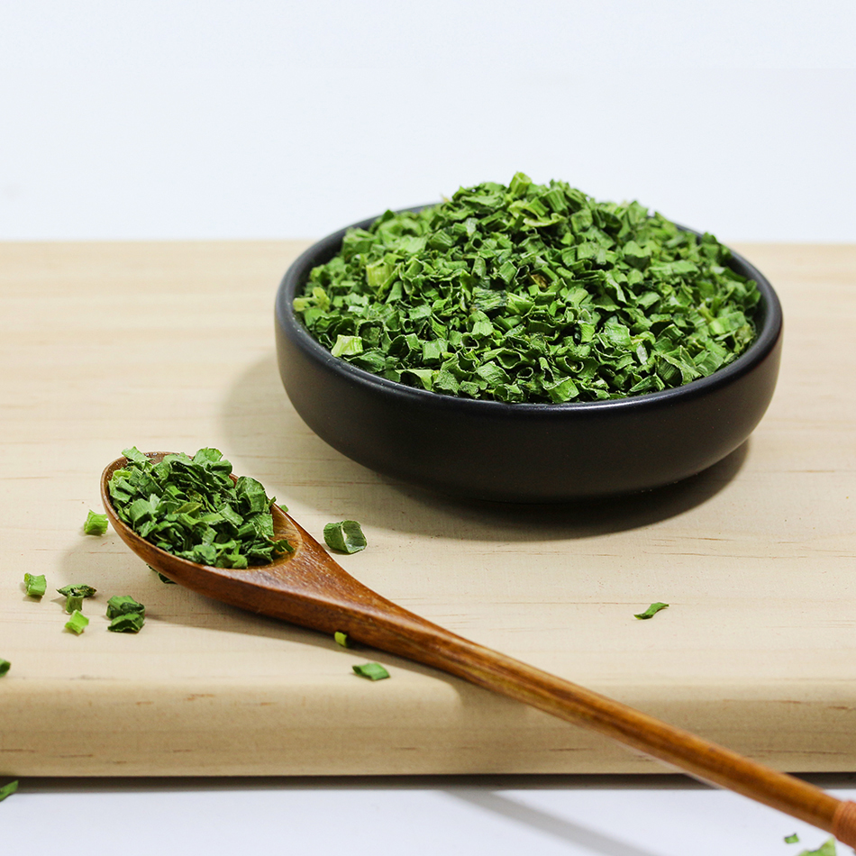
Dehydrated millet onions are made by air-drying fresh millet onions for easy transportation and storage. It is used in family meals, restaurants and seasonings. Delicious and delicious, it is an indispensable spice in life. In life, people don't know how to eat dehydrated millet onions. Here are some practical methods of millet onions. First of all, dehydrated dried onions can stimulate the body's sweat glands and achieve the effect of sweating and heat dissipation. Scallion oil can stimulate the upper respiratory tract, making it easier to cough up sticky phlegm. Onions contain allicin, which has obvious anti-bacterial and viral effects. In particular, the inhibitory effect on Shigella and dermatophytes is more obvious.

Dehydrated Millet Shallot,Millet Shallots For Cooking Noodles,Dehydrated Millet Spring Onion,Delicious Dehydrated Millet Spring Onion
Laian Xinshuyu Food Co., Ltd , https://www.xinshuyufood.com