See how Synaptive Medical subverts neurosurgery
See how Synaptive Medical subverts neurosurgery
June 15, 2015 Source: Health
Window._bd_share_config={ "common":{ "bdSnsKey":{ },"bdText":"","bdMini":"2","bdMiniList":false,"bdPic":"","bdStyle":" 0","bdSize":"16"},"share":{ }};with(document)0[(getElementsByTagName('head')[0]||body).appendChild(createElement('script')) .src='http://bdimg.share.baidu.com/static/api/js/share.js?v=89860593.js?cdnversion='+~(-new Date()/36e5)];It is believed that most neurosurgical doctors have had such experiences. When they are faced with brain charts or models during training, the complex structure can be seen at a glance: the prefrontal cortex stores long-term memory, cerebellar balance, and parietal integration sensory information. However, when the actual craniotomy is performed, it is not the same thing. Only a paste-like group is seen, and the rules that are instantly turned back are completely forgotten. Brain surgery is extremely demanding, and a difference of one millimeter in the lower knife position may permanently damage a patient's hearing, language ability, mobility or sensory ability. Under current conditions, neurosurgeons use models and corpses to learn and train before the actual surgery, which is still far from the brain in actual surgery.
In fact, in addition to doctor training, neurosurgery is relatively backward in technology compared to other medical fields.
In the past half century, the ancient magnifying glass has been the standard optical device in external surgery. The supernatural surgeon either wears a surgical magnifying glass that has been used since 1870, or chooses to mount a large microscope arm above the patient's head. This arm is equipped with a set of binoculars but lacks flexibility. Sometimes, adjusting the arm due to the movement of the surgical site may take a quarter of a time.
In addition to surgical equipment, another obstacle to extra-saccular surgery is the presence of severe faults between various techniques. For example, doctors can use MRI to perform preoperative preparations, and in the surgery, optical tools are used for observation. There is no connection between the two. This makes it difficult for doctors to fully integrate and analyze all information.
However, Canada's SynaptiveMedical is confident that it can solve the pain of these extra-surgical surgeries. Synaptive Medical has invited more than 50 engineers and scientists to work on neurosurgery innovations and eventually developed the BrightMatter range. These include a Brain Simulator, a Dive, a Planner, and a Guide to improve neurosurgery.

Piron, co-founder and president of Synaptive Medical, explained: "In the beginning, we just wanted to improve the imaging problems in the brain during extra-surgical surgery, such as building brain network maps, neuralgrams, etc.. When we go deep into this field, Only after discovering that there are more and more important problems to be solved, we have this series of products." This series of products combines the steps of filming, planning, navigation and adjustment into one, and it is done in one go.
The material of this simulated brain was developed by the Synaptive team of chemists and materials scientists, highly simulating the doctor's sense of the brain tissue in the actual surgery. This kind of simulation is almost weird. This has also filled the gap between doctor training and actual operation, which can improve the accuracy of surgery and minimize brain damage caused by surgery.
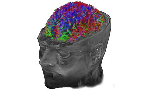
Before the actual surgery, BrightMatter Plan can help doctors plan the operation. High-fidelity magnetic resonance (MR) and diffusion tensor fusion imaging (DTI) are available on the screen to provide a variety of possible brain surgery tracks. This DTI imaging can use the rainbow curve to show the fibers and tissues of the brain. It is easy to see the direction of each tissue and nerve, so that doctors can choose the right position to cut the knife without damaging other parts.
The workflow-driven user interface allows you to design surgical procedures based on images, step by step through 3D imaging and simplified spatial calculations. It can design a variety of surgical procedures in a matter of minutes, measure the selected sulcus pattern, and evaluate the way the skull is cut. With automated data analysis and state-of-the-art 3D imaging, doctors can avoid many tools during surgery and concentrate on surgery.
In the actual operation, BrightMatter Drive replaces the old and bulky microscope arm. The auto-positioning system is also equipped with a microscope, and the robot-like arm automatically moves the position according to the doctor's surgical site, and the image is placed on the large screen by the navigation software to ensure that the scalpel appears on the screen immediately. In addition, the doctor can also control the position through the foot pedal.
MotherBoard's author EreneStergiopoulos exclaimed after experiencing Synaptive's new technology: "Everyone in the operating room can see my every move. Usually, only surgeons can actually see the surgery during surgery. This exclusivity gives the doctor an authority, and under Synaptive's technology, this exclusivity is completely wiped out. However, this also makes the operating room more united."
Even more amazing is that the patient on the operating table can also see the whole process of the operation. In many neurosurgical procedures, the patient is kept awake to detect whether certain operations of the doctor have damaged a certain nerve, and the patient has lost the function of this part. Now, they can listen to a relaxing joke while watching white gloves and scalpels move back and forth in their brains. Moreover, it is their cut brain that controls their senses and nerves to experience this process. It’s really nothing to record your craniotomy. You can also watch the live broadcast.
Laundry Detergent,Organic Detergent Liquid,Laundry Liquid Detergent,Detergent Liquid
Wuxi Keni Daily Cosmetics Co.,Ltd , https://www.kenidailycosmetics.com