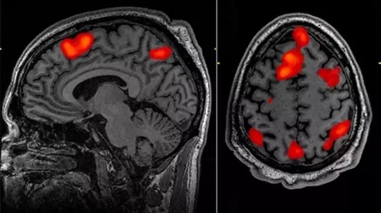
fMRI analysis is plagued by major flaws in brain science
Swedish scientists downloaded a large number of functional magnetic resonance imaging data from brain science and analyzed that fMRI software has a very high probability of false positives when judging brain activity.
It is no exaggeration to say that functional magnetic resonance imaging (fMRI) has brought about earth-shaking changes in the field of neuroscience. When the activity level of different brain regions changes, the blood flow will change accordingly. Neuroscientists use NMR to collect changes in blood flow in each brain region. Using this technology they can non-invasively identify brain regions that are responsible for handling different tasks (such as playing economics games or reading text).
However, this research method and users have received a lot of criticism. Some people worry that this technology exaggerates our ability to read the human mind. Some have pointed out that improper analysis of fMRI data may lead to misleading conclusions, such as a study on dead catfish.

Although the above problems are often caused by poor statistical methods, a study published in the Proceedings of the National Academy of Sciences (PNAS) (Eklund, Anders, Thomas E. Nichols, and Hans Knutsson. “Cluster failure: Why fMRI inferences For spatial extent have inflated false-positive rates. "Proceedings of the National Academy of Sciences (2016): 201602413. The basic information of the paper is at the end of the article.) The problem is much more serious. Some of the basic algorithms involved in fMRI analysis produce false positive "signals" and are of high frequency.
The principle behind fMRI is simple: nerve activity requires energy, and the energy consumed needs to be replenished. This means that blood flow in newly active brain regions will increase. High-resolution MRI can be used to obtain this blood flow data, and the researchers use this to identify brain structures that are activated when a task is performed.
However, the application of this theory in practice is quite complicated. The imaging process divides the brain into tiny three-dimensional units called voxels, and then records the activity in each voxel separately.
Because the voxels are very small, the software must examine the whole and look for "clustering" - a group of neighboring voxels with similar behaviors. The significant result of the dead squid study was that the software was configured by default to be unable to process the huge voxel values ​​of the current MRI scan output. That is to say, even at the 95% confidence level, false positives are inevitable.
Preserved Salted Black Bean,Roasted Salted Broad Beans,Preserved Black Bean With Ginger,Salt And Chilli Edamame Beans
jiangmen city hongsing food co., ltd. , https://www.jmhongsing.com