Indian doctors first use 3D printing technology for tongue cancer surgery
Release date: 2016-08-26

Recently, a group of surgeons from the HealthCare Global Cancer Centre in Bangalore, India, used the 3D printing model to prepare tongue cancer surgery for the first time, making India the world's first 3D printing technology for tongue cancer treatment. country.
The patient was a 53-year-old Indore, Mr. Ravi (a pseudonym). Two years ago, Mr. Ravi received treatment because of a tongue tumor. This time, he had to seek medical treatment again because of the frequent recurrence of oral ulcers. The doctor gave him a magnetic resonance imaging (MRI) scan that showed that Mr. Ravi's tongue tumor had spread over a large area. This is a challenge for the Head & Neck Surgical Oncology team because the large spread of the tumor means that a significant portion of Mr. Ravi's tongue may have to be removed.

Dr. Vishal Rao, the team's head, explained that the number of tongue cancer patients is rising sharply in India. “India is the world's capital of oral cancer. Oral cancer or tongue cancer is quite common in India due to bad habits such as chewing tobacco,†Dr. Rao said. In fact, 40% of cancers in India are oral cancers, and doctors urgently need a low-cost solution to treat the epidemic.
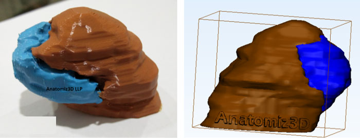
3D printing has proven to be such a low-cost solution because it provides surgeons with information that will help them accurately remove the cancerous parts of the tongue and throat, rather than cutting the entire tongue. Therefore, Dr. Rao and his team turned to the local 3D printing expert Anatomiz 3D LLP. “They made a 3D printed model based on the tongue and the tumor with a simple color division that accurately replicated Mr. Ravi's tongue,†Dr. Rao explained.
To create a 3D model of the tongue and tumor, the Anatomiz3D LLP uses Materialise's Mimics software, which allows them to edit DICOM data from CT scans and MRI scans, so the built 3D model can be printed in a variety of materials and colors. They built a model based on Mr. Ravi's MRI scan, which was divided into healthy parts and cancerous parts. The two parts were printed in different colors with a print ratio of 1:1 and printed materials were a variety of flexible materials. According to Anatomiz3D LLP, the final model is perfect to help surgeons understand the depth, location and size of the tumor.

As shown above (blue represents the cancerous part, brown represents the healthy tissue), the advantages of this method are clearly visible. Because of 3D printing, the surgeon can accurately see the condition of the tongue and plan the removal of the tumor accordingly. This is a huge advancement for oncologists. Their commonly used palpation techniques use 2D images, and 2D images do not adequately show the spread of cancer cells.
The plastic surgery team headed by Dr. Puranik is also very pleased to have such a 3D model. The model provides accurate and necessary data for their tissue implantation surgery, ensuring a satisfactory surgical outcome. Accordingly, Mr. Ravi also benefited from it, so he can retain most of the swallowing and speaking functions. The 3D print model also makes it easier for patients to understand upcoming surgery, and doctors have found that MRI scans are not always understood by patients. This also means that patients can be better prepared to receive surgical results and rehabilitation plans.
Mr. Ravi's surgery has been very successful, and his life is now completely without the shadow of cancer. To the best of our knowledge, this is the world's first tongue cancer surgery using a 3D printed model.
Source: Tiangongshe
Daily Cleaning Nitrile Gloves,Hospital Medical Grade Gloves,Medical Grade Nitrile Gloves Blue,Medical Grade Blue Nitrile Gloves
Puyang Linshi Medical Supplies Co., Ltd. , https://www.linshimedical.com